
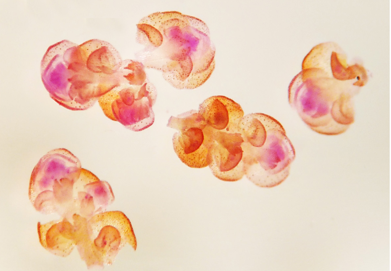

Abstract
Cytogenetic studies in bryophytes have been limited by the difficulty of obtaining sufficient dividing nuclei and by the absence of modern protocols. The technical difficulties stem from the plants’ small size and lack of roots and pollen mother cells, the main sources of cells in division in vascular plants. In bryophytes instead, tiny sporophytes, antheridia, or phyllid meristems must be used to obtain meiotic or mitotic chromosome spreads. We here describe the preparation of such spreads from phyllids, antheridia, and sporophytes in several species of liverworts and compare available protocols with or without prefixation treatments. We also provide illustrated step-by-step instructions. The three prefixation agents (including colchicine) that we tested failed to improve synchronization of cell divisions. Young sporophytes were the best source of diploid synchronized cells, while antheridia were the best source of haploid cells. For meiotic nuclei, a short fixation of capsule tissue at the right developmental stage with 45% acetic acid sufficed to conserve the DNA for cytological investigations, while for mitotic nuclei, fixation in 3:1 ethanol/glacial acetic acid for a longer period (4–24 h) worked well.
Downloads
References
Allen, C.E.A. (1917) chromosome difference correlated with the sex differences in Sphaerocarpos. Science 46: 466–467. https://doi.org/10.1126/science.46.1193.466
Bachtrog, D., Kirkpatrick, M., Mank, J.E., McDaniel, S.F., Pires, J.C., Rice, W.R. & Valenzuela, N. (2011) Are all sex chromosomes created equal? Trends in Genetics 27: 350–357. https://doi.org/10.1016/j.tig.2011.05.005
Berrie, G.K. (1960) The chromosome number of liverworts. The British Bryological Society 3: 688–705. https://doi.org/10.1179/006813860804828963
Brown, R.C. & Lemmon, B.E. (1990) Polar organizers mark division axis prior to preprophase band formation in mitosis of the hepatic Reboulia hemisphaerica (Bryophyta). Protoplasma 156: 74–81. https://doi.org/10.1007/BF01666508
Brown, R.C. & Lemmon, B.E. (2004) γ-tubulin, microtubule arrays, and quadripolarity during sporogenesis in the hepatic Aneura pinguis (Metzgeriales). Journal of Plant Research 117: 371–376. https://doi.org/10.1007/s10265-004-0168-0
Brown, R.C. & Lemmon, B.E. (2011) Spores before sporophytes: hypothesizing the origin of sporogenesis at the algal–plant transition. New Phytologist 190: 875–881. https://doi.org/10.1111/j.1469-8137.2011.03709.x
Brown, R.C., Lemmon, B.E. & Horio, T. (2004) γ-tubulin localization changes from discrete polar organizers to anastral spindles and phragmoplasts in mitosis of Marchantia polymorpha L. Protoplasma 224: 187–193. https://doi.org/10.1007/s00709-004-0061-7
Bull, J.J. (1983) Evolution of sex determining mechanisms. The Benjamin/Cummings Publishing Company, Menlo Park, CA, USA.
Bull, J.J. (1978) Sex chromosomes in haploid dioecy: A unique contrast to Muller’s theory for diploid dioecy. Amer.Naturalist 112: 235–250. https://doi.org/10.1086/283267
Fritsch, R. (1975) Zytologische Untersuchungen an Bryophyten aus dem Harz. Hercynia Neue Folge 12: 75–79.
Heitz, E. (1928) Das Heterochromatin der Moose. I. Jahrbücher für Wissenschaftliche Botanik 69: 762–818.
Iverson, G.B. (1963) Karyotype evolution in the leafy liverwort genus Frullania. The Journal of the Hattori Botanical Laboratory 26: 119–170.
Lorbeer, G. (1934) Die Zytologie der Lebermoose mit besonderer Berücksichtigung allgemeiner Chromosomenfragen. Jahrbücher für Wissenschaftliche Botanik 80: 567–818.
Newton, M.E. (1984) The cytogenetics of bryophytes. In: Dyer, A.F. & Duckett, J.G. (Eds.) The Experimental Biology of Bryophytes. Academic, London, pp. 65–96.
Orzechowska, M., Siwinska, D. & Maluszynska, J. (2010) Molecular cytogenetic analyses of haploid and allopolyploid Pellia species. Journal of Bryology 32: 113–121. https://doi.org/10.1179/037366810X12578498136075
Sone, T., Fujisawa, M., Takenaka, M., Nakagawa, S., Yamaoka, S., Sakaida, M., Nishiyama, R., Yamato, K.T., Ohmido, N., Fukui, K., Fukuzawa, H. & Ohyama, K. (1999) Bryophyte 5S rDNA was inserted into 35S rDNA repeat units after the divergence from higher land plants. Plant Molecular Biology 41: 679–685. https://doi.org/10.1023/A:1006398419556
Sousa, A., Bechteler, J., Temsch, E.M. & Renner, S.S. (2020) Different from tracheophytes, liverworts commonly have mixed 35S and 5S arrays. Annals of Botany 125: 1057–1064. https://doi.org/10.1093/aob/mcaa027
Sousa, A., Schubert, V. & Renner, S.S. (2021) Centromere organization and UU/V sex chromosome behavior in a liverwort. [Online] https://doi.org/10.1111/tpj.15150
Tatuno, S. (1941) Zytologische Untersuchungen über die Lebermoose von Japan. Journal of science of the Hiroshima University. Series B. Division 2. Botany 4: 73–187.
Van Hook, J.M. (1900) Notes on the division of the cell and nucleus in liverworts. Botanical Gazette 30: 394–399. https://doi.org/10.1086/328062
Zheng, M. & Zhu, R-L. (2008) Liverwort anatomy and cytology. Fieldiana 47: 81–87.