Abstract
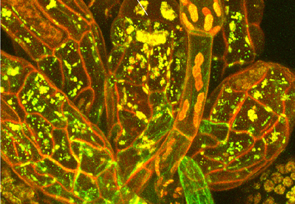
New observations and an overview of the axillary hair structure in the groups of mosses sister to peristomate ones are provided. Three moss lineages, Takakiopsida, Sphagnopsida, and Andreaeobryopsida are characterized by mucilage release through the apical pore or otherwise apical structures. Axillary hairs of Andreaea however, differ from these lineages and are similar to all other mosses, where mucilage exudes through small temporary breakings along the entire cell wall of its upper cells. Fine structure of axillary hairs is illustrated for Sphagnum, Takakia, Andreaeobryum, and partly for Andreaea and Oedipodium, and general characteristics are given for most orders. A unique arrangement of axillary hairs is found in Sphagnum, where they occasionally appear not only in the leaf axils but also along the leaf base abaxially. Another rare feature of Sphagnum is that its axillary hairs are penetrable for fungi through the apical pore, which was previously known in mosses only for Takakia. A highly specialized structure regulating mucilage release in Takakia is described. Actin filaments and especially tubulin microtubules were found to be outstandingly abundant in axillary hairs of Physcomitrium but not in paraphyses, as seen in its GFP-labelled plants, and this feature may be common in other mosses.
Downloads
References
Akiyama, H. (1988) Studies on Leucodon (Leucodontaceae, Musci) and related genera in East Asia. IV. Taxonomic revision of Leucodon in East Asia. Journal of the Hattori Botanical Laboratory 65: 1–80.
Allen, B.H. (2014) Maine mosses: Drummondiaceae–Polytrichaceae. Memoirs of the New York Botanical Garden 111: 1–607.
Allen, B.H. (2018) Moss flora of Central America. Part 4. Fabroniaceae–Polytrichaceae. Monographs in Systematic Botany from the Missouri Botanical Garden 132: 1–830.
Ashton, N.W. & Cove, D.J. (1977) The isolation and preliminary characterization of auxotrophic and analogue resistant mutants of the moss, Physcomitrium patens. Molecular and General Genetic 154: 87–95. https://doi.org/10.1007/BF00265581
Baker, R.G.E. (1988) The morphology and distribution of pits in the cell walls of Sphagnum. Journal of the Hattori Botanical Laboratory 64: 359–365.
Berthier, J., Bonnot, E.-J., Fabre, M.-C. & Hébant, C. (1974) L’appareil sécréteur des Bryales: données morphologiques ultrastructurales et cytochimiques. Bulletin de la Société Botanique de France 121: 97–100. https://doi.org/10.1080/00378941.1974.10839292
Bryan, G.S. (1915). The archegonium of Sphagnum subsecundum. Botanical Gazette 59 (1): 40–56. https://doi.org/10.1086/331467
Bonnot, E.J. & Hébant, C. (1970). Precisions sur la structure et le fonctionnement des cellules mucigenes de Polytrichum juniperinum Willd. Comptes Rendus des Siences de t’ Acadimie des Sciences Serie D, Paris 271: 53–55.
Buck, W.R. (1987) Taxonomic and nomenclatural rearrangements in the Hookeriales with notes on West Indian taxa. Brittonia 39: 210–224. https://doi.org/10.2307/2807378
Buck, W.R. (1988) Another view of familial delimitation in the Hookeriales. Journal of the Hattori Botanical Laboratory 64: 29–36.
Buck, W.R. (1998) Pleurocarpous mosses of the West Indies. Memoirs of the New York Botanic Garden 82: 1–400.
Cox, C.J., Goffinet, B., Neton, A.E., Shaw, A.J. & Hedderson, T.A.J. (2000) Phylogenetic relationships among the diplolepideous-alternate mosses (Bryidae) inferred from nuclear and chloroplast DNA sequences. Bryologist 103: 224–241. https://doi.org/10.1639/0007-2745(2000)103[0224:PRATDA]2.0.CO;2
Crandall-Stotler, B.J. & Bozzola, J.J. (1988) Fine structure of the meristematic cells of Takakia lepidozioides Hatt. et H. Inoue. Journal of the Hattori Botanical Laboratory 64: 197–218.
Duckett, J.G., Carafa, A. & Ligrone, R. (2006) A highly differentiated glomeromycotean association with the mucilage-secreting, primitive Antipodean liverwort Treubia (Treubiaceae): clues to the origins of mycorrhizas. American Journal of Botany 93: 797–813. https://doi.org/10.3732/ajb.93.6.797
Fahn, A. (1979) Secretory tissues in plants. Academic Press, London. New-York. San Francisco.
Goebel, K. (1900) Organographie der Pflanzen, in besondere Archegoniaten und Samenpflanzen, Zweiter Teil: 1–838.
Griffin III, D. (1991) The use of axillary hairs in the taxonomy of two neotropic Bartramiaceae. Journal of Bryology 16: 61–65. https://doi.org/10.1179/jbr.1990.16.1.61
Griffin III, D. (1998) Axillary hairs in Bartramia sect. Strictidium. Evansia 15 (2): 81–83. https://doi.org/10.5962/p.346452
Griffin III, D. & Buck, W.R. (1989) Taxonomic and phylogenetic studies in the Bartramiaceae. The Bryologist 92 (3): 368–380. https://doi.org/10.2307/3243406
Hébant, C. & Bonnot, E.J. (1974) Histochemical studies on the mucilage-secreting hairs of the apex of the leafy gametophyte of some polytrichaceous mosses. Zeitschrift für Pflanzenphysiologie 72 (3): 213–219. https://doi.org/10.1016/S0044-328X(74)80050-4
Hedenäs, L. (1990) Axillary hairs in pleurocarpous mosses a comparative study. Lindbergia 15: 166–180.
Hedwig, J. (1782). Fundamentum Historiae Naturalis Muscorum Frondosorum. 2 Vols. Siegfried Lebrecht, Leipzig. https://doi.org/10.5962/bhl.title.82187
Ignatov, M.S. & Hedenäs, L. (2007) Homologies of stem structures in pleurocarpous mosses, especially of pseudoparaphyllia and similar organs. In: Newton, A.E. & Tangney, R. (Eds.) Pleurocarpous mosses: systematics and evolution. CRC Press, Boca Raton-London-New York (Systematic Association Special Volume 71), pp. 269–286. https://doi.org/10.1201/9781420005592.ch13
Ignatov, M.S., Ignatova, E.A., Fedosov, V.E., Ivanov, O.V., Ivanova, E.I., Kolesnikova, M.A., Polevova, S.V., Spirina, U.N. & Voronkova, T.V. (2016) Andreaeobryum macrosporum (Andreaeobryopsida) in Russia, with additional data on its morphology. Arctoa 25: 1–51. 166–180. https://doi.org/10.15298/arctoa.25.01
Ignatov, M.S., Spirina, U.N., Kolesnikova, M.A., Volosnova, L.F., Polevova, S.V. & Ignatova, E.A. (2018) Buxbaumia: a moss peristome without a peristomial formula. Arctoa 27: 172–202. 166–180. https://doi.org/10.15298/arctoa.27.17
Ignatov, M.S., Spirina, U.N., Kolesnikova, M.A. & Ignatova, E.A. (2021) How opposite may differ from opposite: a lesson from the peristome development in the moss Discelium. Botanical Journal of the Linnean Society 195: 420–436. https://doi.org/10.1093/botlinnean/boaa085
Kozub, D., Khmelik, V., Shapoval, Yu., Chentsov, V., Yatsenko, S., Litovchenko, B. & Starykh, V. (2008) Helicon Focus Software. [http://www.heliconsoft.com]
Landberg, K., Pederson, E.R.A., Viaene, T., Bozorg, B., Friml, J., Jönsson, H., Thelander, M. & Sundberg, E. (2013) The moss Physcomitrium patens reproductive organ development is highly organized, Affected by the Two SHI/STY Genes and by the Level of Active Auxin in theSHI/STY Expression Domain. Plant Physiology 162 (3): 1406–1419. https://doi.org/10.1104/pp.113.214023
Leitgeb, H. (1868) Wachstum des Stammchens von Fontinalis. Sitzungsberichte der Kaiserlichen Akademie der Wissenschaften Mathematisch-Naturwissenschaftlichen Classe 57 (1): 308–342.
Ligrone, R. (1986) Structure, development and cytochemistry of mucilage-secreting hairs in the moss Timiella barbuloides (Brid.) Moenk. Annals of Botany 58 (6): 859–868. https://doi.org/10.1093/oxfordjournals.aob.a087268
Ligrone, R. & Duckett, J.G. (2011) Morphology versus molecules in moss phylogeny: new insights (or controversies) from placental and vascular anatomy in Oedipodium griffithianum. Plant Systematics and Evolution 296: 275–282. https://doi.org/10.1007/s00606-011-0496-1
Liu, Y., Johnson, M.G., Cox, C.J., Medina, R., Devos, N., Vanderpoorten, A., Hedenäs, L., Bell, N., Shevock, J.R., Aguero, B., Quandt, D., Wickett, N., Shaw, A.J. & Goffinet, B. (2019) Resolution of the ordinal phylogeny of mosses using targeted exons from organellar and nuclear genomes. Nature Communications 10: 1485 [1–11]. https://doi.org/10.1038/s41467-019-09454-w
Lorch, W. (1931) Anatomie der Laumboose. In: Linsbauer, Handbuch der Pflanzenanatomie, Abt. II, Teil 2. Bryophyten: 1–359.
Mansouri, K. (2012) Comparative ultrastructure of apical cells and derivatives in bryophytes, with special reference to plasmodesmata. PhD Thesis, Department of Plant Biology, Southern Illinois University, Carbondale, USA. 312 pp. [https://www.proquest.com/openview/ead5e058a9406b8785578683d5e9996b/1?pq-origsite=gscholar&cbl=18750]
Murray, B.M. (1988) Systematics of the Andreaeopsida (Bryophyta): two orders with links to Takakia. Beihefte zur Nova Hedwigia 90: 289–336.
Nishimura, N. (1985) A revision of the genus Ctenidium (Musci). Journal of the Hattori Botanical Laboratory 58: 1–82.
Parihar, N.S. (1962) An introduction to Embryophyta, vol. 1, Bryophyta, ed. 4. Allababad, pp. 1–377.
Piatkowski, B.T. (2015) Axillary hair developmental ultrastructure and mucilage composition in the moss Physcomitrium patens: microscopic and bioinformatic studies. Master of Science Thesis, Department of Plant Biology, Southern Illinois University, Carbondale, USA, 88 pp.
Proskauer, J.M. (1962) Notes on Hepaticae. IV. The Bryologist 65: 213–233. https://doi.org/10.1639/0007-2745(1962)65[213:NOHI]2.0.CO;2
Saidi, Y., Finka, A., Chakhporanian, M., Zrÿd, J.-P., Schaefer, D.G. & Goloubinoff, P. (2005) Controlled expression of recombinant proteins in Physcomitrium patens by a conditional heat-shock promoter: a tool for plant research and biotechnology. Plant Molecular Biology 59: 697–711. https://doi.org/10.1007/s11103-005-0889-z
Saito, K. (1975) A monograph of Japanese Pottiaceae (Musci). Journal of the Hattori Botanical Laboratory 39: 373–537.
Satjarak A., Karen Golinski, G., Trest, M.T. & Graham, L.E. (2022) Microbiome and related structural features of Earth’s most archaic plant indicate early plant symbiosis attributes. Scientific Reports 12: 6423 https://doi.org/10.1038/s41598-022-10186-z
Schimper, W.P. (1858) Versuch einer Entwickelungs-Geschichte der Torfmoose (Sphagnum) und einer Monographie der in Europa vorkommenden Arten dieser Gattung. E. Schweizerbart. 4+96+26 taf., Stuttgart. https://doi.org/10.5962/bhl.title.13447
Schuster, R.M. (1997) On Takakia and the phylogenetic relationships of the Takakiales. Nova Hedwigia 64: 281–310. https://doi.org/10.1127/nova.hedwigia/64/1997/281
Schnepf, E. (1973) Mikrotubulus-Anordnung und -Umordnung, Wandbildung und Zellmorphogenese in jungen Sphagnum-B1ättchen. Protoplasma 78: 145–173. https://doi.org/10.1007/BF01281528
Spirina, U.N., Voronkova T.V. & Ignatov, M.S. (2020) Are all paraphyllia the same? Frontiers in Plant Science 11: 858. https://doi.org/10.3389/fpls.2020.00858
Wanstall, P.J. (1950) Mucilage hairs in Polytrichum. Transactions of the British Bryological Society 1: 349–352. https://doi.org/10.1179/006813850804878798
Whittemore, A.T. & Allen, B.H. (1989) The systematic position of Adelothecium Mitt. and the familial classification of the Hookeriales (Musci). The Bryologist 92: 261–272. https://doi.org/10.2307/3243392
Yamashita, H., Sato, Y., Kanegae, T., Kagawa, T., Wada, M. & Kadota, A. (2011) Chloroplast actin filaments organize meshwork on the photorelocated chloroplasts in the moss Physcomitrium patens. Planta 233: 357–368. https://doi.org/10.1007/s00425-010-1299-2
Verdus, M.C. & Bonnot, E.J. (1982) Secretion mucigene chez Campylopus introflexus (Hedw.) Brid. (Bryopsida). Bulletin de la Société botanique de France, Actualitis botaniques 129 (1): 47–51. https://doi.org/10.1080/01811789.1982.10826548
Zolotov, V.I. & Ignatov, M.S. (2001) On the axillary hairs of Leptobryum (Meesiaceae, Musci) and some other acrocarpous mosses. Arctoa 10: 189–200. https://doi.org/10.15298/arctoa.10.20

