

Abstract
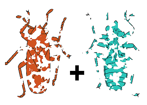
While examining rich insect collections from the Lower Permian Chekarda locality in the Cis-Urals region of Russia, preserved at the Paleontological Institute (PIN) in Moscow, I noticed that dark patches, representing fragments of fossilized cuticle upon a lighter rock matrix, on the part and the counterpart of one fossil often appeared complementary and could be fitted together like a jigsaw puzzle. The phenomenon is well familiar to those who study thin compression fossils. Insects, for example, occasionally become compressed by sediment to such a state that the multiple cuticular layers of their bodies—opposite body sides, overlapping wings, legs, and other appendages—all become fused into a single film of fossilized cuticle. Then, a blow of the rock hammer fractures this film in such a way that some of its fragments become exposed upon the part and others upon the counterpart. I attempted to combine such part and counterpart images into more complete composites and in this way I was able to interpret many poorly preserved specimens, which otherwise would be left out of the study, and, moreover, to see and understand morphological structures of crucial importance, which otherwise would be overlooked. The results of my palaeoentomological study will be published elsewhere, but my success in superposition of part and counterpart images warrants a separate, brief publication on the method itself. Undoubtedly the idea must have occurred to many. Merged composites of part and counterpart images and descriptions of the implemented merging techniques have been published as part of larger studies (e.g., Haug et al., 2009; Haug & Haug, 2013). Yet, I believe the method deserves a dedicated discussion. It has some limitations, which confine the scope of its use, but in some cases it may help visualize poorly preserved, badly fragmented specimens, which may otherwise remain uninterpretable. Therefore, the goal of the present note is to describe a simple method of part-counterpart (PCP) image merging and discuss its pitfalls and limitations. The images were processed manually using either of two computer graphics editors, the proprietary Adobe Photoshop and the free open-source GIMP. In this form, the technique is available to virtually anyone. On the other hand, its computer automation appears to be a realistic goal.
References
- Adobe Photoshop Help. Available from: https://helpx.adobe.com/pdf/photoshop_reference.pdf (Accessed 28 May 2025).
- GIMP User Manual. Available from: https://docs.gimp.org/2.10/en/ (Accessed 28 May 2025).
- Haug, C., Haug, J.T., Waloszek, D., Maas, A., Frattigiani, R. & Liebau, S. (2009) New methods to document fossils from lithographic limestones of southern Germany and Lebanon. Palaeontologia Electronica, 12 (3), 6T, 12 pp.
- Haug, J.T. & Haug, C. (2013) An unusual fossil larva, the ontogeny of achelatan lobsters, and the evolution of metamorphosis. Bulletin of Geosciences, 88, 195–206. https://doi.org/10.3140/bull.geosci.1374
- Jepson, J.E., Ansorge, J. & Jarzembowski, E.A. (2011) New snakeflies (Insecta: Raphidioptera) from the Lower Cretaceous of the UK, Spain and Brazil. Palaeontology, 54 (2), 385–395. https://doi.org/10.1111/j.1475-4983.2011.01038.x