
Abstract
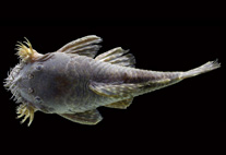
The species of Rhinoderma Duméril & Bibron are endemic to the temperate forests of South America in southern Chile and Argentina (Formas et al. 1975). Both have specialized reproductive modes, Rhinoderma darwinii Duméril & Bibron undergoes complete embryonic and larval development in the mouth of the male: newly metamorphosed frogs are expelled into the terrestrial environment (Jorquera et al. 1972). In contrast, embryos of R. rufum (Philippi) do not remain in the male's mouth, but instead are expelled into the water as larvae (Jorquera et al. 1974). Jorquera et al. (1972, 1974) described the normal development of both species. The chondrocranial morphology and visceral skeleton of R. darwinii was described by Lavilla (1987) and its internal oral features by Wassersug & Heyer (1988). The table of normal development of R. rufum (Jorquera et al. 1974) emphasized the duration of each stage of development: however, some features currently used for comparative purposes in tadpole morphological studies were not included or described only briefly. In this work, I include a detailed description of the morphology, particularly of the mouthparts, of the tadpoles of R. rufum, and describe the chondrocranium, hyobranchial skeleton, and the internal features of the oral cavity in this species. In addition, I compare the morphology of the mouth, chondrocranium, and hyobranchial skeleton of the congeneric tadpoles, considering different modes of feeding (endotrophic in R. darwinii versus exotrophic in R. rufum). Last, I present some thoughts about the evolution of larvae of Rhinoderma.